- Printed Journal
- Indexed Journal
- Refereed Journal
- Peer Reviewed Journal
P-ISSN: 2349-6800, E-ISSN: 2320-7078


Journal of Entomology and Zoology Studies
2019, Vol. 7, Issue 6
Gross and morphometrical studies on the humerus of Indian Barking deer (Muntiacus muntjak)
Kamal Sarma, Jasvinder Singh Sasan and Shalini Suri
The present study was conducted on the humerus of an adult Indian Barking deer. It was a long bone with spirally twisted shaft and two prominent extremities. The proximal third of the medial surface had a short teres tubercle. The proximal third of the lateral surface possessed at its middle a sharp deltoid tuberosity. The nutrient foramen was located on the distal third of the shaft on the postero-lateral aspect. The head was elliptical in outline with a very distinct neck. The cranial part or summit of lateral tuberosity was well developed and blunt whereas the caudal part was ill-developed. The medial surface of the summit of lateral tuberosity facing the bicipital groove had 2 spine-like structures, the distal of which was better developed than the proximal one. The area for the insertion of infra-spinatus muscle was roughly triangular in outline. The medial tuberosity was much smaller as compared to the lateral one. A distinct groove was observed caudal to the posterior division of the medial tuberosity. The bicipital or inter-tubercular groove was well developed and roughly U-shaped. The anterior parts of both the lateral and medial tuberosities curved over the bicipital groove. The distal extremity consisted of two condyles, two epicondyles and two fossae. The medial condyle and epicondyle were much larger than the lateral counterpart but the lateral epicondylar crest was more prominent than the medial one. The radial fossa was deep but the olecranon fossa was much deeper. Both the fossae were separated by a thin plate of bone. The width of the bone decreased from proximal to the middle of the shaft and then increased towards distal end.
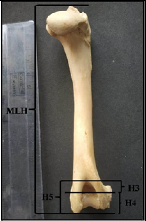
Fig. 1: Photograph showing lengths of different segments of humerus of Indian Barking deer
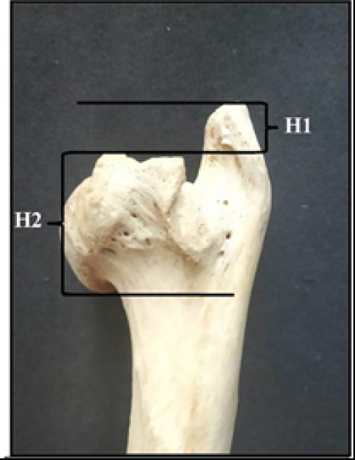
Fig. 2: Photograph showing lengths of different segments of humerus of Indian Barking deer (contd.)
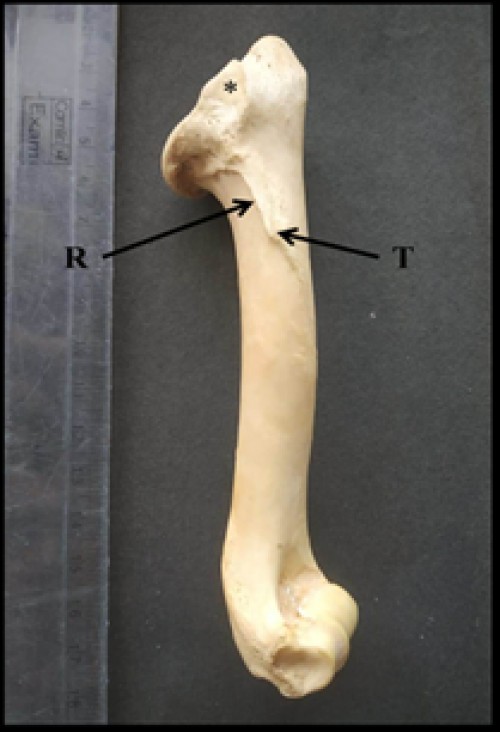
Fig. 3: Photograph showing lateral surface of humerus of Indian Barking deer showing triangular shaped area for insertion of infra-spinatus muscle (*), deltoid ridge (R) and deltoid tuberosity (T)
Pages : 278-282 | 721 Views | 115 Downloads


How to cite this article:
Kamal Sarma, Jasvinder Singh Sasan, Shalini Suri. Gross and morphometrical studies on the humerus of Indian Barking deer (Muntiacus muntjak). J Entomol Zool Stud 2019;7(6):278-282.
Indexed In

Hard Copy Subscription
Click Here for more InformationOur Other Journal

Important Topics
Related Links
Related Journal Subscription
Important Publications Links
Important Links

Journal of Entomology and Zoology Studies

- Home
- Editorial Board
- Archives
- Instructions
- Membership
- Publication Ethics
- Publish Book (ISBN)
- Make Payment
- Contact Us
- Helpline No.: +91-9711224068
- Fast Publication: +91-7048922346
- Toll Free: 1800-1234070
