- Printed Journal
- Indexed Journal
- Refereed Journal
- Peer Reviewed Journal
P-ISSN: 2349-6800, E-ISSN: 2320-7078


Journal of Entomology and Zoology Studies
2019, Vol. 7, Issue 6
Gross and morphometrical studies on the humerus of Indian Barking deer (Muntiacus muntjak)
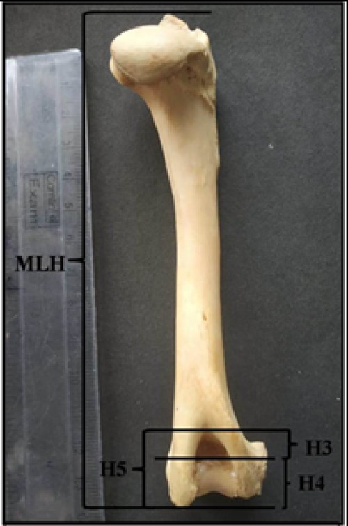
Fig. 1: Photograph showing lengths of different segments of humerus of Indian Barking deer
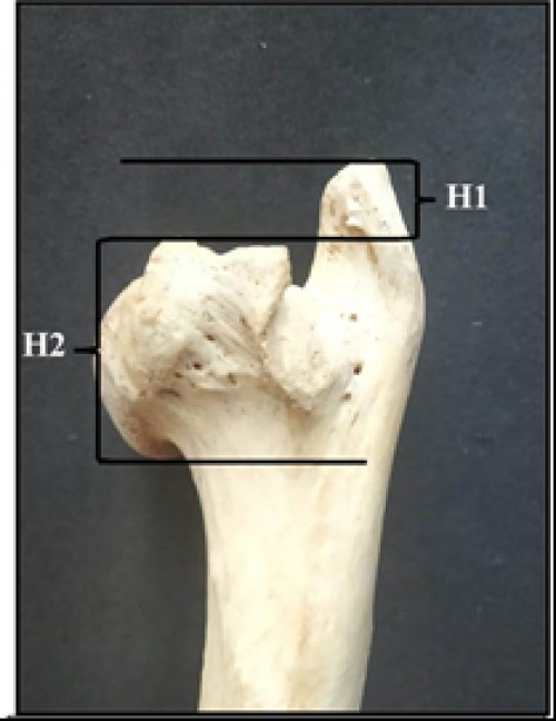
Fig. 2: Photograph showing lengths of different segments of humerus of Indian Barking deer (contd.)
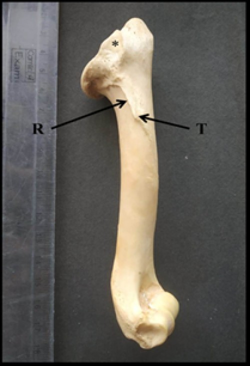
Fig. 3: Photograph showing lateral surface of humerus of Indian Barking deer showing triangular shaped area for insertion of infra-spinatus muscle (*), deltoid ridge (R) and deltoid tuberosity (T)
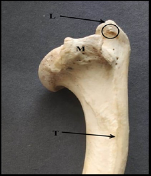
Fig. 4: Photograph showing medial surface of humerus of Indian Barking deer showing teres tubercle (T), medial tuberosity (M), lateral tuberosity (L) and two spine-like structures on lateral tuberosity (encircled)

Fig. 5: Photograph showing anterior surface of humerus of Indian Barking deer showing medial tuberosity (M) and lateral tuberosity (L) curving over the bicipital groove (B), radial fossa (R), medial condyle (C1) and lateral condyle (C2)
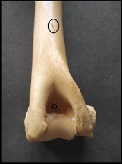
Fig. 6: Photograph showing distal extremity of humerus of Indian Barking deer showing nutrient foramen (encircled) and deep olecranon fossa (O)
Indexed In

Hard Copy Subscription
Click Here for more InformationOur Other Journal

Important Topics
Related Links
Related Journal Subscription
Important Publications Links
Important Links

Journal of Entomology and Zoology Studies

- Home
- Editorial Board
- Archives
- Instructions
- Membership
- Publication Ethics
- Publish Book (ISBN)
- Make Payment
- Contact Us
- Helpline No.: +91-9711224068
- Fast Publication: +91-7048922346
- Toll Free: 1800-1234070
